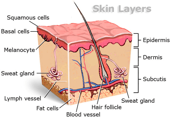
What is the epidermis? We can define the epidermis as one or more layers of cells forming the tough and protective outer layer of the skin or integument (natural coating). The epidermis is a protective outer covering of many plants and animals. It may be comprised of a single layer, as in plants, or of several layers of cells on top of the dermis, as in those of vertebrate animals.

Figure 1: skin anatomy and layers – diagram. Credit: Mark Kelly, Heartstring.net
There are five layers of the epidermis. These are the stratum basale, the stratum spinosum, the stratum granulosum, the stratum lucidum, and the stratum corneum. The stratum lucidum is found only in thick skin and is located between the stratum corneum and the stratum granulosum. Figure 2 shows the layers of the epidermis.
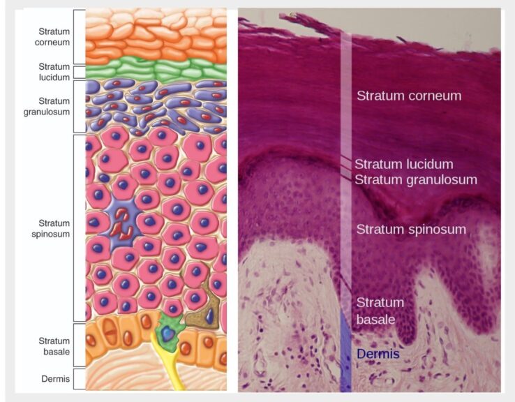
In humans, the skin is the largest organ of the integumentary system. It is composed of two major layers: (1) epidermis and (2) dermis. The epidermis is the outer, waterproofed layer of the skin and the dermis is the layer below the epidermis. In between the two layers is a thin sheet of fibers called the basement membrane.
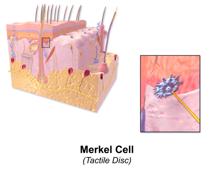
Other cells found in the stratum basale are Merkel cells (Figure 4). Merkel cells were originally discovered by Friedrich Merkel in the late 1800s. They are scarce, specialized epithelial cells found directly above the basement membrane and function as type-1 mechano-receptors. They can sense light/gentle touch as their membrane interacts with nerve endings found in the skin. Therefore, they are found mainly in lips, fingertips, and the face where sensory perception is at its most sensitive. On the other side of their membrane, they are linked to keratinocytes by desmosome multiprotein complexes.
The stratum basale is attached to the basement membrane by desmosomes and hemidesmosomes. These are multiprotein complexes allowing strong cell adhesion. They are intercellular junctions that give mechanical strength to the tissue.
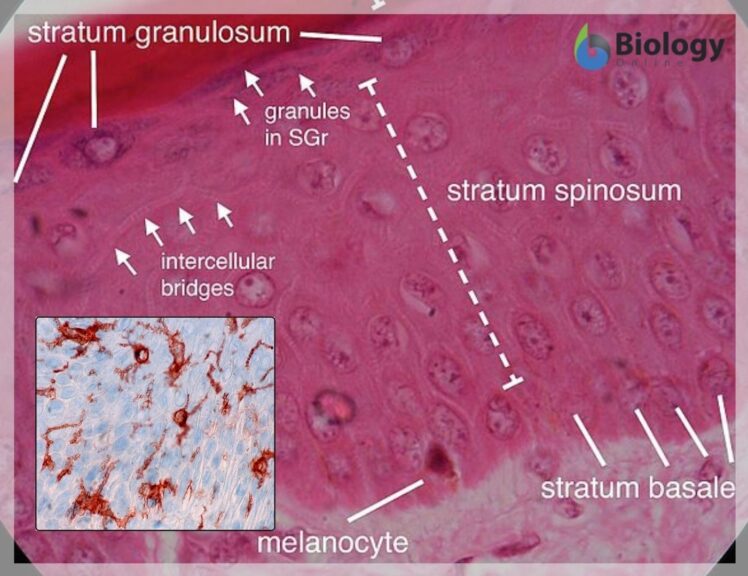
The layer above the stratum basale is the stratum spinosum, also known as the prickle layer. It is around 8 – 10 cells thick and it contains polyhedral shaped cells. The cells in this layer contain many desmosomes which function to anchor the cells together. When this cell layer is prepared on a slide for microscopy, the spaces between the desmosomes shrink away giving the cells a spiky appearance. This spiky appearance is not how the cells appear in real life. The desmosomes act as structural support and provide flexibility and elasticity.
Dendritic cells of the immune system are also found in the superficial portion of this layer. Dendritic cells are termed Langerhans cells when they are found in the epidermis. They lie very close to keratinocytes in this layer. They also extend their processes to the next layer, the stratum granulosum. These Langerhans cells recognize pathogens and allergens in the epidermal layers and migrate to the lymph node where they can initiate an appropriate immune response.
As new keratinocytes move into this layer, they push older keratinocytes out into the next epidermal layer of the skin, the stratum granulosum.
In humans and other vertebrates, the epidermis consists of the following layers (strata):
The epidermis has many functions including acting as a water barrier, structural stability, immune defense, body homeostasis, endocrine and exocrine functions, and sensation/touch. Each will be discussed in more detail below.
Sweat glands on the other hand are eccrine glands that respond to body temperature. These are found in large quantities in skin tissue throughout the body, particularly the palms of hands and soles of the feet. Their job is to secrete water and electrolytes which disperse through the epidermis allowing the regulation of body temperature through sweating.
As mentioned earlier, Merkel cells are involved in the mediation of the perception of light touch. Merkel cells are described as neuroendocrine cells or mechanoreceptors. They are found in clusters in areas called touch domes. Free nerve endings are also found in the epidermis that aid with sensory functions including sensations to heat, touch, cold, and pain.
The role of the skin is vital as it protects the body (especially the underlying tissues) against pathogens and excessive water loss. It is also involved in providing insulation, temperature regulation and sensation.
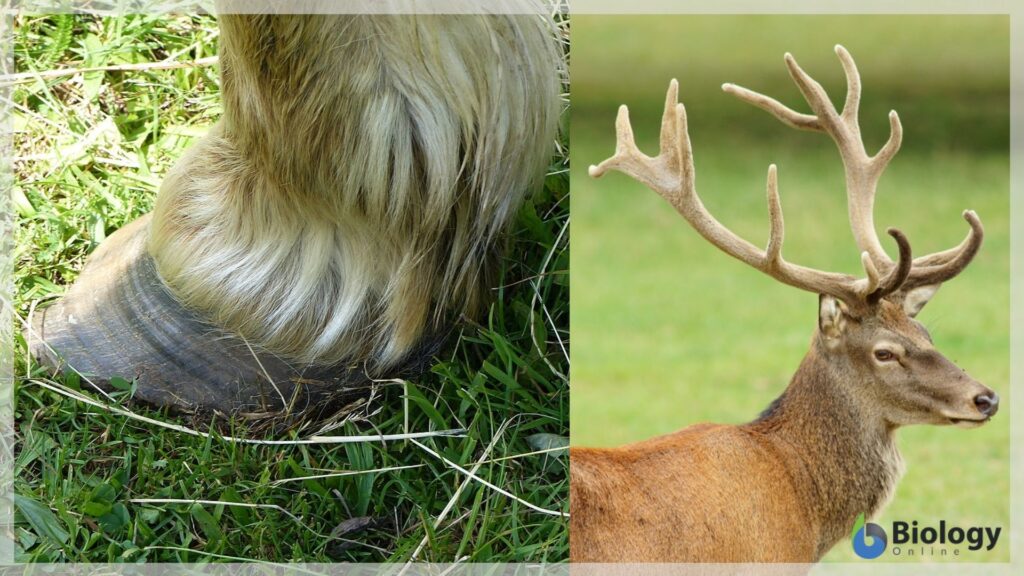
Figure 7: hoof (left) and antlers (right). Source: Modified by Maria Victoria Gonzaga, BiologyOnline.com, from the works of Tsaag Valren (hoof photo), CC BY-SA 4.0 and Peter Trimming, CC BY 2.0.
Hooves, nails, claws, and horns are some of the toughest biological materials due to the strength of the dead keratin-filled cells. Due to the complex nature of keratinous material, their formation can serve a wide range of biological properties such as impact resistance (as in hooves), external attack (horns and nails), and withstanding aerodynamic forces (feathers).
The epidermis covers all herbaceous plants and is found covering the leaves, stems, flowers, and roots. Above ground, the epidermal cells of plants have a waxy coating known as a cuticle which is impermeable to water. (Figure 9) The cuticle keeps water in and pathogens out providing a protective role. An epicuticular wax covers the epidermis of some plants. This further protects plants from water loss as well as wind and strong sunlight. These plants are adapted to living in hot, dry environments. The cuticle is transparent allowing light to go through it. There are little to no chloroplasts in the epidermal layer, these are found in the layers below and as the epidermis is translucent, the sunlight can reach them with ease.

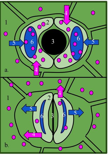
Stomata are present in the epidermis of leaves, these are openings that allow gas exchange. The stomata are made up of a pair of guard cells that make the stomatal pore. These guard cells contain chloroplasts allowing them to make food using photosynthesis. (Figure 10)
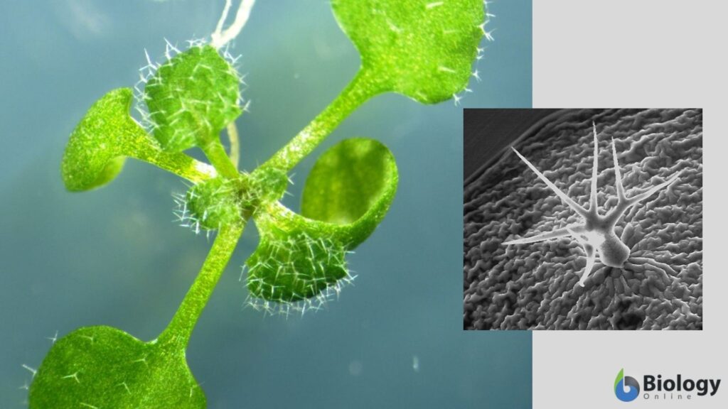
Extensions of the epidermis can be seen in most plants, these are called trichomes that act to protect the plant from insects or herbivores. They can do this by either secreting toxic substances or by preventing pests from reaching the plant surface (spikes or spines). Stinging nettles are one such example where their trichomes can break off and inject the animal/human with histamines that irritate. In insectivorous plants, as in the sundews (Drosera), sticky nectar is produced by the trichomes and this attracts and catches insects for the plant to digest.
The epidermis is also found in the roots of plants. Here, it acts to allow the uptake of water up the root system. The root epidermis has a very thin cuticle to allow water uptake. Roots may produce a chemical called mucigel, which is a carbohydrate that is hydrophilic allowing the root to move through the soil easily.
In plants, the epidermis is a single layer of cells as opposed to the several layers of cells in the human and animal epidermis. The plant epidermis also has an additional layer on top of it — the cuticle, which is an impervious substance secreted by epidermal cells to protect against desiccation (water loss).
The epidermis is present in animals and plants as an outer protective layer providing a vital barrier to environmental pathogens, chemicals, and UV as well as having an important structural role. In animals, the epidermis has been adapted to individual species to provide protection, defense, and body regulation by the formation of hooves, hairs, feathers, and nails. Similarly, plants have adapted to their environment by either increasing the thickness of their cuticle on the epidermis depending on wet or dry environments. They also have a wide range of trichomes that act to deter pests and herbivores. Overall, the epidermis is accountable for the safety of the organism in which it covers.
Try to answer the quiz below to check what you have learned so far about the epidermis.